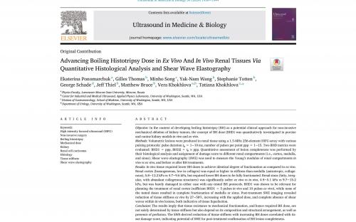
The results of our study on the mechanical dose of renal tissue ablation have been published in the Q1 journal Ultrasound in Medicine and Biology
Boiling histotripsy method that is being developed at LIMU can potentially be applied for non-invasive mechanical fractionation of various biological tissues, including, for example, renal tumors. However, kidneys consist of compartments that differ in structure and mechanical properties - and, accordingly, in sensitivity to histotripsy:
- cortex – external part, homogeneous, with low collagen content;
- medulla – middle part, anisotropic, collagenous;
- sinus – center of the kidney, fatty, irregular, with abundant collagenous structures.
In addition, renal tumors themselves can also differ in their structure, properties, and degree of perfusion (i.e., supply of fluid and, in particular, blood).
Thus, the paper was aimed at quantitative studies of:
- stiffness of various kidney compartments in vivo (i.e., inside the body, with active blood flow) and ex vivo (i.e., outside the body, in the absence of blood flow) using shear wave elastography;
- mechanical dose required for successful fractionation of these kidney compartments using boiling histotripsy method.
For quantitative analysis of the susceptibility of different kidney compartments to varied mechanical doses, a specific scale of mechanical damage was developed for each kidney compartment based on microscopic photographs of histological sections of the kidneys containing the lesions.
It was shown that the tissue resistance to mechanical fractionation, and hence required histotripsy dose, are not solely determined by tissue stiffness but also depend on:
- the amount of collagen: higher collagen content increases tissue resistance to histotripsy;
- the degree of perfusion: the presence of blood/fluid facilitates the histotripsy treatment.
In the future, these results will be useful in pre-treatment planning for fractionation of various renal tumors depending on their composition, structure and degree of perfusion.
For more details – see the text of the paper.


