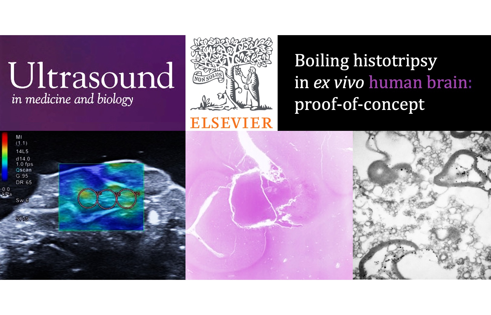
Our results on histotripsy fractionation of human brain tissues ex vivo have been published in the Q1 journal Ultrasound in Medicine and Biology


Currently, the standard method for radical treatment of brain diseases is surgical intervention to remove certain areas of the brain (e.g., tumors), which involves skull trepanation (opening the cranium) and further incision through healthy tissues on the way to the target area. Such intervention is associated with the risk of bleeding, infections and other postoperative complications, and the outcome largely depends on the size of the target area and its localization in the brain.
Currently actively developing methods of non-invasive surgery using high-intensity focused ultrasound (HIFU) allow for non-invasive ablation of specified areas in the brain by ultrasonic heating of tissue up to the temperatures of thermal necrosis under MRI control. Thermal HIFU technology is currently used in clinics in Russia and other countries to treat movement disorders (e.g., essential tremor, tremor in Parkinson's disease, dystonia, etc.) by generating localized thermal ablation inside the brain.
Despite the clinical success of transcranial HIFU surgery, its application is yet limited to the central parts of the brain - thalamus and globus pallidus - due to the risk of overheating the skull bones that effectively absorb ultrasound during shallow focusing. Other limiting factors are heat diffusion and cooling of the heated area by blood flow, which reduce the accuracy of the ablation, as well as the need for expensive MR thermography devices to monitor temperature during surgery.
Boiling histotripsy (BH) is a new developing HIFU method that allows for non-invasive mechanical fractionation of unwanted tissues in the human body (e.g., tumors) using short HIFU pulses with shock fronts at the focus. Rapid heating of the tissue at the focus leads to its boiling within milliseconds and formation of vapor cavities and cavitation microbubbles. Their interaction with the ultrasound shock fronts leads to mechanical fractionation (liquefication) of the tissue into subcellular fragments. Nonthermal mechanism of the method can minimize the limitations of the existing thermal HIFU approach and expand the spectrum of clinical applications addressed with focused ultrasound. Specifically, BH allows for visualization of the procedure using conventional diagnostic ultrasound: vapor inclusions in tissue during tissue fractionation appear bright on ultrasound, whereas the liquefied tissue appears dark.
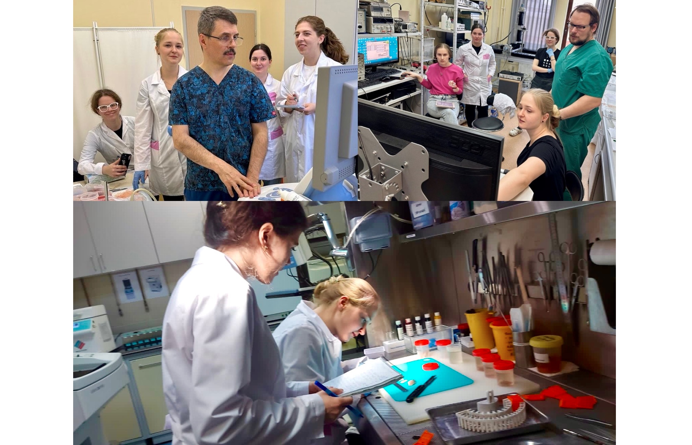
Numerical experiments performed by our lab have previously shown the possibility of implementing the conditions and safe use of BH in brain tissue transcranially, including close to the skull. However, the ability to mechanically fractionate human brain tissue using this method has not yet been experimentally studied. The results of the studies published in our paper in collaboration with Moscow State University Clinic, Pulmonology Scientific Research Institute, and Research Institute of Human Morphology were primarily devoted to experimental demonstration of the possibility of BH fractionation of human brain tissues – to begin with, ex vivo (i.e., outside the body) and in the absence of the skull.
BH fractionation of various human brain tissues (grey matter, white matter, thalamus and globus pallidus) was carried out under ultrasound imaging guidance with varied exposure parameters. The resulting damage was analyzed at the macroscopic level (through lesion bisection and photographing), microscopic level (histologically), and ultrastructural level (by transmission and scanning electron microscopy). All experiments were accompanied by preliminary measurements of the tissue stiffness using shear wave elastography, since susceptibility to histotripsy is mainly determined by elastic properties of the target tissue.
In the published studies, feasibility of BH for fractionation of various human brain tissues ex vivo into subcellular fragments was demonstrated for the first time. The fractionation process was accelerated by the use of shorter pulses with larger amplitudes without loss of efficiency. It was also shown that stiffer white matter, mainly consisting of nerve fibers, is more difficult to fractionate than softer grey matter, consisting of neuron bodies. In addition, for the first time it was possible to generate mechanical damage in thalamus and globus pallidus - those parts of the brain that in modern clinical practice are thermally ablated with HIFU to treat movement disorders under rather expensive MRI control.
Our further studies will be aimed at experimental demonstration of BH feasibility in the presence of skull on the ultrasound beam path to bring the laboratory experiments closer to clinical conditions. Further successful implementation of BH into clinical practice will potentially reduce the number of open brain surgeries and the cost of such interventions due to the possibility of non-invasive fractionation of the target areas of the brain under conventional diagnostic ultrasound imaging.
For more details – see the text of the paper.