Our joint study with the Moscow State University Clinic is the first to demonstrate the feasibility of non-thermal ultrasound-guided fractionation of leiomyosarcoma ex vivo (i.e., outside the body) using boiling histotripsy under diagnostic ultrasound guidance. The results have been published in a top-rated journal Ultrasonics (Q1, IF 4.1) in the paper titled “Non-thermal ultrasound-guided fractionation of human leiomyosarcoma with boiling histotripsy: an ex vivo feasibility study“.
Leiomyosarcoma is an aggressive and rapidly growing malignant tumor that develops from smooth muscle tissue and is one of the most common types of sarcomas. This tumor can arise in various anatomical sites, but is most commonly found in the retroperitoneum, uterus, extremities, and blood vessels. Currently, the standard treatment is surgical removal, sometimes combined with radiation and chemotherapy. However, not all patients can tolerate this treatment, and inoperable cases are treated only with palliative care. Therefore, researchers are searching for new gentle yet effective methods for local non-surgical treatment of leiomyosarcoma.
The idea behind boiling histotripsy lies in focusing a periodic sequence of high-amplitude millisecond-long ultrasound pulses from a transducer located outside the patient’s body through the skin and healthy tissue onto the pathological area. This area is destroyed not by ultrasonic heating but mechanically through the action of nonlinear acoustic waves with sharp pressure changes (shock fronts) on the tumor. As a result, the unwanted tissue is remotely “blended” into a structureless suspension, without damaging the intervening healthy tissues or surrounding the target area. The entire process can be observed and monitored using widely available diagnostic ultrasound equipment.
In previous studies, we have demonstrated that the tissue resistance to mechanical fractionation with ultrasound (histotripsy) is largely determined not only by the tissue stiffness but also by its composition and structural arrangement. Specifically, heterogeneous tissues with distinct structural patterns, such as myocardium, uterine fibroids, renal medulla, and brain white matter, are more resistant to mechanical fractionation with ultrasound and, therefore, require higher doses for complete fractionation than homogeneous tissues of equal or even higher stiffness. Since leiomyosarcoma is primarily composed of smooth muscle tissue (similar to, for example, uterine fibroids, which are difficult to mechanically fractionate), its resistance to histotripsy and the corresponding required doses were investigated in the published study.
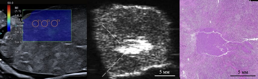
In our joint study with physicians from the Moscow State University Clinic, we used autopsy specimens of human renal and retroperitoneal leiomyosarcoma. Their clinical relevance was preliminarily assessed using a diagnostic ultrasound system with shear wave elastography based on the stiffness of the autopsy specimens compared to the clinically observed values. The specimens were then placed into a histotripsy system specifically developed by our group for laboratory experiments and containing a focused ultrasound transducer. Short high-intensity ultrasound pulses of 1–2 ms duration were focused inside the tumor to mechanically fractionate the planned volumes of malignant tissue under diagnostic ultrasound guidance.
Histological analysis of the tumor microstructure after histotripsy treatment confirmed complete fractionation of the tumor cells in the target volumes while maintaining clear boundaries of less than 200 microns between the destroyed and intact tissue. Furthermore, the duration of each procedure fractionating the planned volumes of malignant tissue up to 230 cubic mm did not exceed 20 minutes and was successfully controlled using conventional ultrasound imaging. Further optimization of ultrasound parameters will reduce the procedure time, making it even more attractive for clinical use as a non-invasive alternative to existing invasive and minimally invasive treatments for leiomyosarcoma.
In our work, a classic leiomyosarcoma was studied which is the most commonly found subtype of this tumor. It can be hypothesized that other types of leiomyosarcomas, such as myxoid leiomyosarcomas with a lower cell density or pleomorphic leiomyosarcomas with a lower connective tissue content, will be even more sensitive to mechanical fractionation with histotripsy. Our future research will focus on optimizing ultrasound protocols for various types of leiomyosarcomas, adapting the method for clinical use taking into account ultrasound beam aberrations when passing through skin and soft tissue, and studying the potential immunological effects of histotripsy in the treatment of inoperable sarcomas.
The obtained results demonstrate that leiomyosarcoma can be effectively mechanically fractionated with high-amplitude pulsed focused ultrasound and lay a solid foundation for future in vivo studies. This opens the way for the development of non-invasive methods for the local treatment of sarcomas, especially in cases where surgery is not possible.
For more details – see the text of the paper.